SDB Volunteers Shine at
5th USA Science &
Engineering Festival
By Marsha E. Lucas
The Society for Developmental Biology along with
BioEYES participated in the
5th USA Science &
Engineering Festival held April 6-8, 2018 in
Washington, D.C. Our exhibit “Development, Growth,
and Regeneration” featured developing fish and frog
embryos and highlighted how diverse animals
including zebrafish and planaria are able to regrow lost body parts. More than 50 volunteers
supported our exhibit over the three-day festival.
|
 |
|
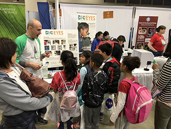
Steve Farber and his captive
audience. Credit: Christine Weston |
BioEYES co-founder, Steve Farber of the Carnegie
Institution for Science, provided zebrafish and many microscopes
for visitors to observe development from a single
cell through larval stages. He and volunteers from
BioEYES, the Carnegie and Johns Hopkins
University helped visitors identify male and female
adult zebrafish, determine the stage of embryos, and observe
circulating blood and a beating heart.
|
 |
|
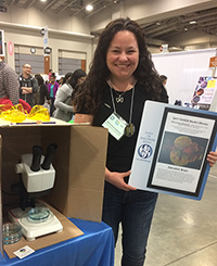
Marina Venero Galanternik with
her FASEB BioArt winning image. Credit:
Marsha Lucas |
Marina Venero Galanternik,
a postdoc in
Brant Weinstein's lab at the National Institutes
of Health, excited attendees with her GFP-expressing zebrafish.
She won the
2017 FASEB BioArt
contest for an image of the vasculature surrounding
the brain in these same fish.
Himani Datta Majumdar from
Sally Moody’s lab at
George Washington University provided Xenopus
embryos and tadpoles. Kids and adults learned about
the lifecycle of frogs, identified the developmental
stage of the embryos and tadpoles, and observed the
beating heart.
This was the first year SDB had an exhibit on
regeneration. Visitors learned about the importance
of stem cells in regeneration and that diverse
organisms undergo this process.
|
 |
|
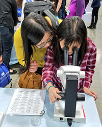
Mom and daughter observe Xenopus
embryos. Credit: Carnegie Institution
for Science |
At the
planaria station, guests observed tail and head
fragments in the process of regeneration. They
identified the site where new (white) tissue was
growing and learned how that tissue contained stem
cells that will form many different cell types.
At
the zebrafish regeneration station, clear glasses
housed individual adult fish with cut or uncut tail
fins. Again visitors were able to observe white
tissue at the amputation site and see how after
three weeks the tail fin was indistinguishable from
an uncut tail fin.
Marnie Halpern of the Carnegie
Institution for Science and members of her lab photographed the
regeneration process over three weeks and created a
beautiful vertical banner for the exhibit.
|
 |
|
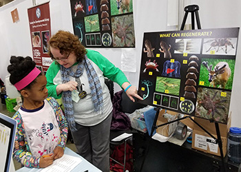
Marnie Halpern assists student in
regeneration game. Credit:
Christine Weston |
Halpern also designed a “What can regenerate?” game
in which visitors had to identify the pictured
organisms that regenerate. Kids received some cool SDB
swag for their efforts.
We thank all of the students, postdocs, faculty and
friends of the Society for Developmental who
traveled from across the Mid-Atlantic region to
volunteer their time, materials, and expertise
toward the success of this science festival. Also, a
special thanks to Voot Yin of MDI Biological
Laboratory and Kate McDole of HHMI-Janelia for their
beautiful movies.
Check out the SDB website for
more photos from the festival!
|