2013 BSDB Spring Meeting Report
By John Young
 John
Young won the best student poster competition at the
Society for Developmental Biology's
71st Annual Meeting in Montréal, Canada which sent
him to the 2013 British Society for Developmental
Biology's spring meeting in Conventry, England.
The following is Young's report from the meeting. John
Young won the best student poster competition at the
Society for Developmental Biology's
71st Annual Meeting in Montréal, Canada which sent
him to the 2013 British Society for Developmental
Biology's spring meeting in Conventry, England.
The following is Young's report from the meeting.
I'm John Young, a
graduate student at the University of California,
Berkeley in
Richard Harland's lab. I'm interested in
axial patterning of the vertebrate embryo.
Specifically, I've been studying the role of
canonical Wnt signaling in posteriorization of the
neural plate in the frog Xenopus. I'm
grateful to SDB for sending me to attend this
meeting as part of the student poster award from the
2012 meeting in Montréal, Quebec.
The 2013
British
Society for Developmental Biology/Cell Biology joint
meeting was held on the campus of the University of
Warwick in Coventry, England. Given that the meeting
included both of these societies, the talks and
posters presented topics that ranged from the actin
dynamics of sub-cellular structures to the genetics
of left-right patterning. The science was exciting,
the people welcoming, and England was a wonderful
place to visit, even in March!
The meeting began
unofficially with a graduate student symposium where
six students were selected to present 15-minute
talks on the poster that they would present. An
excellent idea, it was a nice introduction to the
breadth of topics that would be covered during the
meeting in a less formal setting that encouraged
questions from fellow students as well as lead
investigators. Stephen Fleenor (2012 BSDB best
student poster winner from
Jo Begbie's lab) talked
about his identification of specific splice forms of
the signaling regulator RGS3 and how these isoforms
mediate the transition between progenitor and
differentiating neurons. Also in this session, Siyao
Wang (Gino Poulin's lab) presented her findings that
depletion of the MLL/COMPASS complex, responsible
for DNA methylation, results in somatic cells with
germ cell like characteristics.
Olivier Pourquié
gave the first of two opening plenary talks on the
mechanisms of segmentation in the vertebrate axis.
Work from his group has shown that many signaling
pathways and cyclically expressed genes regulate
this process. He showed data from microarray
analyses his group did on discrete samples of
dissected somitic mesoderm along the anterior
posterior axis to understand how cyclic gene
expression is different. The data resolved into four
clusters corresponding to their axial position,
which unexpectedly showed very different metabolic
signatures, providing a novel method of regulation.
David Drubin provided the second plenary talk on
actin dynamics and endocytic vesicle trafficking.
His group engineered fluorescent fusion proteins
expressed at physiological levels in mammalian cells
with zinc-finger nuclease technology. His talk had
one of the most memorable moments of the meeting
when the entire audience collectively gasped when,
using live cell imaging, he showed that clatherin
mediated endocytosis in mammals was indeed a regular
and efficient process, contrary to previously held
beliefs.
|
 |
|
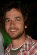
John Young at the Tower of London |
Various concurrent
sessions were presented during the day. In the
epithelia and mechanosensory session,
Shigenobu
Yonemura and
Guillaume Charras highlighted the use
of atomic force microscopy for assaying force
detection in epithelial sheets. Daniel Grimes
(Dominic Norris' lab) presented a nice story on the
function of the polycystin proteins PKD2 and PKD1L1
in left-right patterning of the mouse by using a
novel PDK1L1 gain of function allele, RKS. During
the cancer models session,
David Adams gave an
update of the efforts of the Wellcome Trust in
generating knockout mouse lines and their protocols
in assessing the associated phenotypes. In the gene
regulation session,
Sarah Bray spoke on the multiple
roles of Notch signaling in the differentiation of
blood cell types in Drosophila, and
Patrick Lemaire spoke on the evolution of cis-regulatory
elements governing neural gene expression in
different Ciona species.
Elly Tanaka showed that
axolotls regenerate their limbs via differentiation
of resident stem cells while newts accomplish this
by dedifferentiating muscle cells.
This year's student
poster award winner was
Aditya Saxena from
Helen Skaer's lab at Cambridge. His work identified novel
factors that interact with Rho and Rac to mediate
the morphogenetic movements of cells comprising the
Malpighian tubules of Drosophila. Be sure to
look for his poster at this year's SDB meeting in
Cancun.
The final session
included a talk by
Kathryn Anderson presenting her
lab's finding that mutations in the kinesin Kif7
result in mouse embryos with open neural tubes.
However, this is not due to a loss of the primary
cilium but because of defective cilia that have
overgrown. She showed that Kif7's role is to sever
microtubules of the cilium in order to maintain
proper length and stability.
Robb Krumlauf gave the
final talk of the meeting on the evolution of neural
segmentation. Lamprey eels do not show segmental hox
gene expression in the hindbrain however, he showed
that inserting zebrafish hox gene enhancers into
lampreys resulted in segmental expression.
Personally, the
highlight of the meeting for me was the awards
session on the second night.
Eric Miska received the
Hooke Medal for his work on non-coding RNAs and
their roles in epigenetic regulation of the genome.
Finally, the
Waddington Medal (kept secret until
it's given) was awarded to
Jim Smith who recapped
his work on mesoderm induction and patterning in
Xenopus. It was a particularly inspiring talk to me,
as I am completing my grad work on frogs. Probably
the biggest surprise was when Jim thanked
Sir John
Gurdon who happened to be sitting in the audience
unannounced!
In closing, this was an exciting meeting full of
great talks, posters and people. I certainly hope
the tradition between SDB and BSDB of sending the
poster award winners to each other's annual meetings
continues.
|